top of page

Deformities

Amniotic Band Syndrome (ABS)

Amniotic Band Syndrome (ABS) is an uncommon congenital abnormality that causes entrapment of fetal parts (usually a limb or digits) in fibrous amniotic bands while in utero. It can result in disfigured feet. ABS affects about one in every 1,200 births and is believed to be the cause of 178 in 10,000 miscarriages. Some researchers believe that ABS is caused by early amniotic rupture, which leads to the formation of fibrous strands that entangle limbs and appendages. Treatment may include surgical excision of the fibrous band and any necrotic tissue.

Please contact our office to discuss your specific diagnosis and treatment options.

Claw Toe

Claw toe is caused by nerve damage from diseases like diabetes or alcoholism, which can weaken muscles in the foot. The term stems from the toes' appearance—toes that look like claws digging down into the soles. Claw toe may lead to the formation of painful calluses. Claw toe worsens without treatment and may become a permanent deformity over time. 


Common symptoms of claw toe include:

  • Toes bent upward from the joints at the ball of the foot.

  • Toes bent downward at the middle joints toward the sole of the foot.

  • Corns on the top of the toe or under the ball of the foot.


Claw toe deformities are easier to repair when detected early. Splints or tape is used to hold the toes in correct position.

Club Foot

Clubfoot is one of the most common, non-life threatening, major birth defects among infants globally. Approximately one in every 1,000 newborns has clubfoot. Of those, one in three have both feet clubbed. The exact cause is unknown. Two out of three clubfoot babies are boys. Clubfoot is twice as likely to occur if one or both parents and/or a sibling has had it. Less severe infant foot problems are often incorrectly called clubfoot.


Clubfoot twists the heel and toes inward. It often appears like the top of the foot is on the bottom. Additionally, the clubfoot, calf, and leg are smaller and shorter than normal. When clubfoot is detected at birth, it is not painful and is correctable.


The goal of treating clubfoot is to make the infant's clubfoot (or feet) functional, painless, and stable by the time he or she is ready to walk. Serial casting is the process used to slowly move the bones of a clubfoot into the proper alignment. The chiropodist starts by gently stretching the child's clubfoot toward the correct position. A cast is put on to hold the foot in place. One week later, the cast is removed, the baby's foot is stretched a little farther toward the correct position, and a new cast is applied. X-rays are used throughout the process to check on progress toward proper foot alignment. Casting generally repeats for 6-12 weeks, and may take up to 4 months.


About half the time, clubfoot straightens with casting. Once the proper foot alignment is achieved, the child is fitted with special shoes or braces to keep the foot straight once corrected. These maintenance devices are used until the child has been walking for up to a year or more. Muscles for children with clubfoot commonly try to return to the clubfoot position; a regular occurrence among 2 and 3 year olds, but a condition that may continue up to age 7.


In some cases, stretching, casting, and bracing is not enough to correct clubfoot. Surgery may be required to adjust the tendons, ligaments, and joints in the foot and ankle.

Dysplasia

Dysplasia, also known as epiphysealis hemimelica, is a disorder that affects the bone joints. It is characterized by overgrowth of the cartilage on the end of one or more of the long bones (carpal or tarsal bones) in the hand or foot. Usually only one limb is involved. Dysplasia may cause limbs that are unequal in length. Less often, the cartilage on other bones, such as those in the ankle, knee, or hip joint, can be impacted.

Enchondroma

Enchondromas are small benign tumors made up of cartilage that form in the bone beneath the toenail. Enchondromas are the most common bone tumors of the hands and feet and usually are painless. The tumor can involve large portions of the bones, causing thinning of the cortex. This can weaken the bone and cause it to break spontaneously. When enchondromas occur in the small bone in the end of the toe, they can cause pain that may mimic the pain of ingrown toenails.


Ollier's Disease, also known as enchondromatosis, frequently occurs in the small bones in the hands and toes (phalanges) and the long bones behind the phalanges, called metatarsals. This condition is characterized by multiple enchondromas.


Maffucci's Syndrome is a very rare form of enchondromatosis that combines multiple enchondromas in bones anywhere in the body with benign soft tissue tumors (known as hemangiomas), which are associated with blood vessels. This condition tends to appear in the hands and feet, and has a greater tendency toward malignant transformation than Ollier's Disease.


Because they are painless, most enchondromas are discovered when X-rays are taken for another reason. CT scans and MRI can also help in diagnosing enchondromas.


The majority of enchondromas require no treatment. Only in cases where the tumors are aggressive and begin destroying bone tissue do they require further attention, often surgical removal.

Flat Feet

Gordon Syndrome

Gordon Syndrome is an extremely rare disorder that belongs to a group of genetic disorders known as the distal arthrogryposes. These disorders typically involve stiffness and impaired mobility of certain joints of the lower arms and legs (distal extremities) including the knees, elbows, wrists, and/or ankles. 


Joints affected by this disorder tend to be permanently fixed in a bent or flexed position. In the foot, Gordon Syndrome is characterized by the abnormal bending inward of the foot. The range and severity of symptoms may vary from case to case. Gordon Syndrome is believed to be an inherited condition.

Haglund's Deformity 

Haglund's Deformity (also known as pump bump or retrocalcaneal bursitis) is a painful enlargement on the back of the heel bone that becomes irritated by shoes. It normally appears as a red, painful, and swollen area in the back of the heel bone. Women tend to develop the condition more than men because of irritation from rigid heel counters in shoes (the piece of leather forming the back of a shoe) that rub up and down on the back of the heel bone.


Changing shoes, soaking feet, and anti-inflammatory medications often mitigate the symptoms of this problem. Note: Please consult your physician before taking any medications.

Hallux Limitus

Hallux Limitus is a condition that results in stiffness of the big toe joint. It is normally caused by an abnormal alignment of the long bone behind the big toe joint, called the first metatarsal bone. Left untreated, Hallux Limitus can cause other joint problems, calluses, and/or diabetic foot ulcers. Painful bone spurs also can develop on the top of the big toe joint. 


Anti-inflammatory medications, cortisone injections, and/or functional orthotics are some of the common treatments for stiff big toe. Note: Please consult your physician before taking any medications. Surgery may be required if spurring around the joint becomes severe.

Hammertoes

Jackson-Weiss Syndrome 

Jackson-Weiss Syndrome (JWS) is a rare genetic disorder characterized by foot abnormalities. Symptoms include abnormally broad big toes, webbing of the skin between the second and third toes, an inward angling of the toes, and/or malformation or fusion of certain bones within the feet. Jackson-Weiss Syndrome is inherited and affects both sexes equally.

Mallet Toes

Mallet toes are deformities caused by bone and muscle imbalances that become exaggerated in people with active lifestyles. Arthritis can also lead to mallet toes. Mallet toes can cause extreme discomfort and may be aggravated if restrictive or improperly fitting footwear is worn for a prolonged period of time. 


Treatment is designed to relieve pressure, reduce friction, and transfer forces from the sensitive areas. Shoes with a high and broad toe box (toe area) are recommended to prevent future irritation for people suffering from mallet toes. Other conservative treatments include forefoot supports, such as gel toe caps, gel toe shields, and toe crests. Gel forefoot supports provide immediate comfort and relief from common forefoot disorders without drying the skin.

Metatarsalgia

Pain in the area between the arch and toes, or ball of the foot, is generally called metatarsalgia. The pain usually centers on one or more of the five bones (metatarsals) in this mid-portion of the foot. Also known as dropped metatarsal heads, metatarsalgia can cause abnormal weight distribution due to overpronation.


Metatarsalgia occurs when one of the metatarsal joints becomes painful or inflamed. People often develop a callus under the affected joint. Metatarsalgia also can be caused by arthritis, foot injury (from sports, a car accident, or repeated stress), hard surfaces (cement or tile floors), and specific footwear (rigid-soled work boots).


A simple change of shoes may solve the problem. In more severe cases, custom orthotics may be prescribed to alleviate the pain and prevent overpronation.

Osteomyelitis (Bone Infection)

Osteomyelitis is a type of bacterial bone infection that moves from acute to chronic phases quickly. The infection usually begins in another part of the body and spreads to the bone via blood. Traumatic injury, frequent medication injections, the use of a prosthetic device, and some surgical procedures can increase susceptibility to the underlying infection.


With osteomyelitis, the infected bone fills with a pus that deprives the bone of its needed blood supply. Over time, this can result in the death of bone tissue.


The presence of bone infection can be diagnosed with tests, such as bone scans and MRI.


Osteomyelitis infections are very difficult to cure with oral or intravenous antibiotics. In chronic cases, surgical removal of the dead bone tissue is usually required.

Overlapping or Underlapping Toes

Overlapping toes are characterized by one toe lying on top of an adjacent toe. The fifth toe is the most commonly affected. Overlapping toes may develop in the unborn fetus. Passive stretching and adhesive taping is most commonly used to correct overlapping toes in infants, but the deformity usually recurs. Sometimes they can be surgically corrected by releasing the tendon and soft tissues about the joint at the base of the fifth toe. In some extreme cases, a pin may be surgically inserted to hold the toe in a straightened position. The pin, which exits the tip of the toe, may be left in place for up to three weeks.


Underlapping toes usually involve the fourth and fifth toes. (A special form of underlapping toes is called congenital curly toes). The cause of underlapping toes is unknown. It is speculated that they may be caused by an imbalance in muscle strength of the small muscles of the foot.  If deformed toes are flexible, a simple release of the tendon in the bottom of the toe will allow for them to straighten. If the deformity is rigid, surgery may be needed to remove a small portion of the bone in the toe.

Peroneal Tendon Dislocation

Peroneal tendons support two important foot muscles (Peroneus Brevis and Peroneus Longus) that originate on the outside of the calves. These two muscles allow you to roll to the outside of your foot while standing.


Peroneal tendons are also called stirrup tendons because they help hold up the arch of the foot. The two muscles are held in place by a band of tissue, called the peroneal retinaculum. Injury to the retinaculum can cause this tissue to stretch or tear. When this happens, the peroneal tendons can dislocate from their groove on the back of the fibula. The tendons can be seen to roll over the outside of the fibula, which damages the tendons.


Skiing, football, basketball, and soccer are the most common sports activities leading to peroneal tendon dislocation. In some cases, ankle sprains also have caused this condition. Patients usually have to use crutches after such an injury, in order to allow the retinaculum tissue to heal and the tendons to move back to their natural position on the fibula. Sometimes a splint or compression bandage is applied to decrease swelling. Anti-inflammatory medications and ice are often part of the treatment. Note: Please consult your physician before taking any medications.


In moderate to severe cases of injury, when the peroneal retinaculum is torn or severely stretched and susceptible to dislocation, surgery may be required.

Posterior Tibial Dysfunction

The posterior tibial tendon starts in the calf, stretches down behind the inside of the ankle, and attaches to bones in the middle of the foot. This tendon helps hold the arch up and provides support when stepping off on your toes when walking. If it becomes inflamed, over-stretched or torn, it can cause pain from the inner ankle. Over time, it can lead to losses in the inner arch on the bottom of your foot and result in adult-acquired flatfoot.


Signs and symptoms of posterior tibial tendon dysfunction include:

  • Gradually developing pain on the outer side of the ankle or foot.

  • Loss of the arch and the development of a flatfoot.

  • Pain and swelling on the inside of the ankle.

  • Tenderness over the midfoot, especially when under stress during activity.

  • Weakness and an inability to stand on the toes.

People who are diabetic, overweight, or hypertensive are particularly at risk. X-rays, ultrasound, or MRI may be used to diagnose this condition.


Left untreated, posterior tibial tendon dysfunction may lead to flatfoot and arthritis in the hindfoot. Pain can increase and spread to the outer side of the ankle.


Treatment includes rest, over-the-counter nonsteroidal anti-inflammatory drugs, and immobilization of the foot for six to eight weeks with a rigid below-knee cast or boot to prevent overuse. Note: Please consult your physician before taking any medications.

Sesamoiditis

Sesamoids are small bones that are connected only to tendons or are embedded in muscle. This structure appears in only a few places in the human body, one of which is the foot. Two very small sesamoids (about the size of a kernel of corn) are found in the underside of the forefoot near the big toe—one on the outer side of the foot and the other closer to the middle of the foot. Sesamoids provide a smooth surface over which the tendons slide, thus increasing the ability of the tendons to transmit muscle forces. In the foot, sesamoids assist with weight-bearing and help elevate the bones of the big toe.


Like other bones, sesamoids can fracture. Additionally, the tendons surrounding the sesamoids can become irritated or inflamed, which is called sesamoiditis and is a form of tendonitis. Sesamoiditis is a common condition among ballet dancers, runners, and baseball catchers because of the pressures placed on their feet.


Symptoms include:

  • Pain under the big toe or on the ball of the foot.

  • Swelling and bruising.

  • Difficulty and pain in bending and straightening the big toe.

Surgery is usually not required to treat sesamoiditis. Treatments generally include:

  • Discontinuation of the activity causing the pain and inflammation.

  • Over-the-counter pain medications and anti-inflammatories, such as ibuprofen and aspirin. Note: Please consult your physician before taking any medications.

  • Icing the sole of the foot.

  • Wearing shoes that are soft-soled and low-heeled.

  • Using cushioning in shoes to relieve stress.

  • Injection of a steroidal medication to reduce swelling.

If symptoms persist, you may need to wear a removable brace on the leg for 4 - 6 weeks to give the inflammation time to subside and the bones to heal.

Spurs

Bone spurs are a very common foot problem. In the feet, they develop most frequently in the heel, near the toes, and on top of the big toe joint. The spurs are small outgrowths of bone. In and of themselves, they are generally harmless. However, their location may cause friction or irritation from shoes or other foot structures, which can lead to other foot problems.


Heel spurs refer specifically to bone spurs in the heel. Heel spurs are growths of bone on the underside, forepart of the heel bone and occur when the plantar fibrous band pulls at its attachment to the heel bone. This area of the heel later calcifies to form a spur. With proper warm-up and the use of appropriate athletic shoes, strain to the ligament can be reduced.
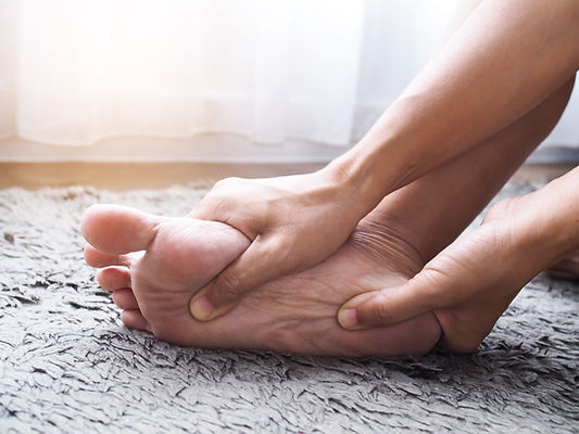
 

Anti-inflammatory medications, cortisone injections, corrective shoes, and/or orthotics (special shoe inserts) are some of the common treatments for spurs. Note: Please consult your physician before taking any medication. Surgery may be prescribed if spurring around the joint becomes severe or leads to recurrent pain from persistent corns.

Tarsal Coalition

Tarsal coalition is a bone condition that causes decreased motion or absence of motion in one or more of the joints in the foot. The bones found at the top of the arch, the heel, and the ankle are referred to as the tarsal bones. A tarsal coalition is an abnormal connection between two of the tarsal bones in the back of the foot or the arch. This abnormal connection between two bones is most commonly an inherited trait.


The lack of motion or absence of motion experienced in a tarsal coalition is caused by abnormal bone, cartilage, or fibrous tissue growth across a joint. When excess bone has grown across a joint, it may result in restricted or a complete lack of motion in that joint. Cartilage or fibrous tissue growth can restrict motion of the affected joint to varying degrees, causing pain in the affected joint and/or in surrounding joints.


Symptoms usually include an aching sensation deep in the foot near the ankle or arch, accompanied by muscle spasms on the outside of the affected leg. Nonsurgical treatments, such as corrective shoes or custom orthotics, physical therapy, and anti-inflammatory medication, are the first courses of action. Note: Please consult your physician before taking any medications. Surgery is sometimes performed in severe cases to allow for more normal motion between the bones or to fuse the affected joint or surrounding joints.

bottom of page